It is a radiographic examination of urinary tract including renal parenchyma, calyces and pelvis after intravenous injection of contrast media or dye into the bloodstream.
Intravenous urography (IVU) also referred as intravenous pyelography (IVP) as it implies the visualization of the pelvis and calyces without the parenchyma.
The term “pyelogram” is reserved for the retrograde studies visualizing only the collecting system.
Anatomy of urinary system
Urinary system refers to the structures that produce and conduct urine to the point of excretion.
COMPONENTS OF THE URINARY SYSTEM
- Two kidneys
Two ureters
Urinary bladder
Urethra
After filter the blood, the remaining water (urine), passes through the ureters and then stored in the urinary bladder.

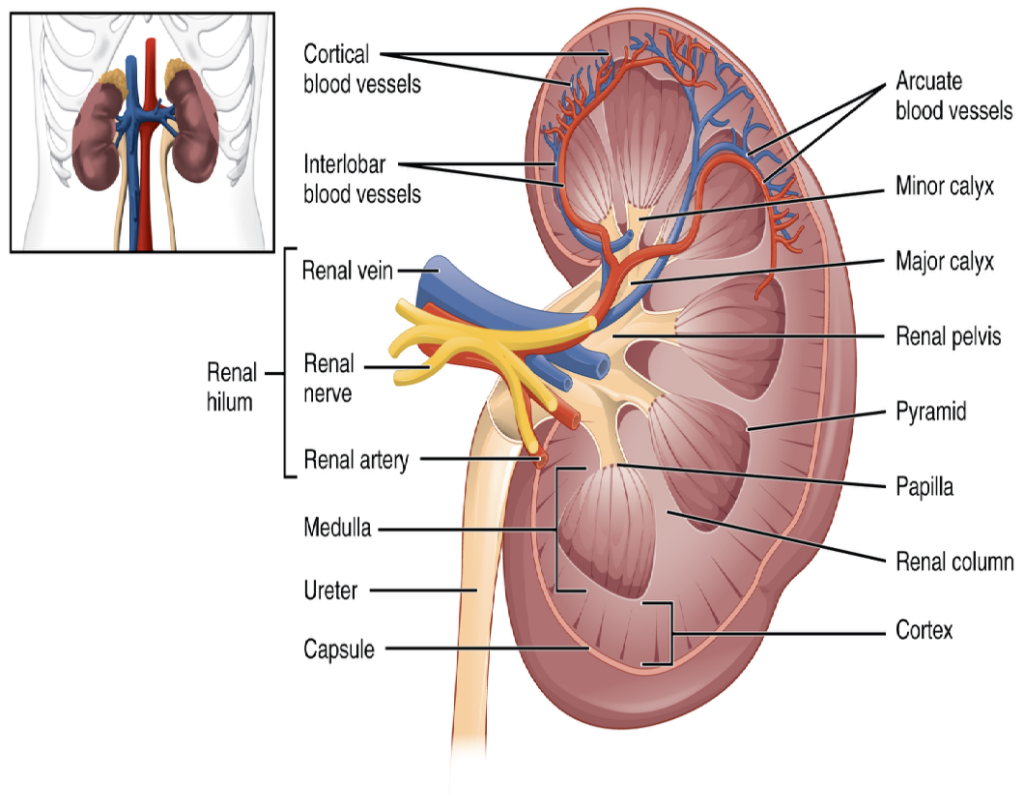
GROSS ANATOMY OF KIDNEY
- The functional unit of the kidney is nephron.
It has two parts: renal parenchyma and renal sinus.
The renal parenchyma of the kidney is divided into two major structures: superficially renal cortex and deep is the renal medulla.
The medulla consists of 6-18 renal pyramids.
Between the renal pyramids are projection of cortex called renal columns.
The papilla or tip, of each pyramid empties urine into a minor calyx.
minor calyces empty into major calyces empty into the renal pelvis, which becomes the ureter.
Function of the urinary system
- The kidneys are bean-shaped organs. They filter blood to remove extra water and waste in urine (wee). Most of us have two kidneys. They are on either side of our spine (backbone), near the bottom edge of our ribs at the back.
The two ureters are long tubes that carry urine from the kidneys to the bladder.
The bladder is a bag that stores urine until we are ready to urinate. It sits low down in the pelvis.
The urethra is a tube that carries urine from the bladder to the outside of the body.
Indications
- Hematuria
To evaluate the ureteral obstruction.
Abnormalities of the ureter.
Differentiation of function of both kidney.
Suspected renal injury.
Renal colic or flank pain.
Urinary tract infection (UTI).
Calculus disease
Contraindication
- Iodine sensitivity
Pregnancy
Previous allergy to contrast agent.
Cardiac and renal failure.
Raised serum creatinine level.
Patient preparation
- Bowel preparation:
Laxatives is recommended to eliminate faecal matter from the colon and to reduce amount of gas in the bowel.
Dulcolax (biscodyl) is given 2-4 tablets at bedtime for 2 days prior to the IVU.
Castor oil is an effective laxative given about 80ml the night before IVU. Castor oil is contraindicated in case of abdominal pain of unknown cause, old and debilitated patients.
Medication required for old age patient and fatty patient. - Low residue diet and plenty of oral fluid is required.
- Ask for any history of diabetes, renal disease, allergy to drugs and any specific food.
Blood creatinine level should be in its normal range (Males- 0.6 to 1.5mg/dl, Females- 0.5-1.5mg/dl)
Blood urea level should range between 9 to 42mg/dl.
Fasting for 4 hours.
Do not dehydrate the patients if patient suffering from renal failure as it may lead to severe fluid and electrolyte imbalance.
Sensitivity to the dye checked. Necessary precautions are taken to avoid the allergic reactions.
Take informed consent.
Extra renal routes for contrast excretion:
- Hepatic
Saliva
Sweat
Tears
Gastric juice
Procedure requirements
- Compression band is applied over distal ends of ureters to retard flow of opacified urine into bladder and to ensure adequate filling of renal pelvis and calyces, so it is required for the procedure.
IV cannula or butterfly needle(20-24G).
Fixing tape
Syringe- 50ml,10ml
Medical Gloves
Tourniquet
Cotton swap
Dry cotton
Emergency drugs
Contrast media
Adult dose – 50-100ml (w/v)
Paediatric dose – 1ml/kg